肛門挙筋
肛門挙筋(こうもんきょきん)は骨盤底筋の一つで、骨盤隔膜を構成し、骨盤内臓を支持し、肛門の周囲に位置する骨筋である恥骨直腸筋・恥骨尾骨筋・腸骨尾骨筋の総称をいう。
肛門挙筋の1つ腸骨尾骨筋(ちょうこつびきん)は、腸骨からおこり尾骨と仙骨につく骨格筋である。
ギャラリー
-
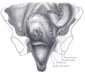
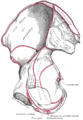 Right hip bone. Internal surface.
 -
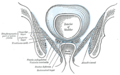 Coronal section of pelvis, showing arrangement of fasciæ. Viewed from behind.
 -
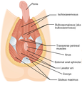 Muscles of male perineum.
 -

-
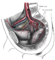 The arteries of the pelvis.
 -
 Sacral plexus of the right side.
 -
 Iliac colon, sigmoid or pelvic colon, and rectum seen from the front, after removal of pubic bones and bladder.
 -
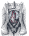 The posterior aspect of the rectum exposed by removing the lower part of the sacrum and the coccyx.
 -
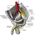
 Male pelvic organs seen from right side.
 -
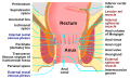 Anatomy of the human anus.


参考文献
この記事にはパブリックドメインであるグレイ解剖学第20版(1918年)422ページ本文が含まれています。
出典
外部リンク
- 『肛門挙筋』 - コトバンク
- Anatomy figure: 41:05-00 at Human Anatomy Online, SUNY Downstate Medical Center —「女性の浅会陰隙の筋肉」。
- Anatomy figure: 42:04-00 at Human Anatomy Online, SUNY Downstate Medical Center —「男性の浅会陰隙の筋肉」。
- Anatomy photo:43:16-0102 at the SUNY Downstate Medical Center —「骨盤横隔膜の筋肉」
- Anatomy image:9072 at the SUNY Downstate Medical Center
- Anatomy image:9089 at the SUNY Downstate Medical Center
- Anatomy image:9871 at the SUNY Downstate Medical Center
- Cross section image: pelvis/pelvis-e12-15ウィーン医科大学のプラスティネーション研究所
- perineum at The Anatomy Lesson by Wesley Norman (Georgetown University)( analtriangle3 )
- pelvis at The Anatomy Lesson by Wesley Norman (Georgetown University)( femalepelvicdiaphragm 、 malepelvicdiaphragm )
- 肛門挙筋症候群に関するメルクのマニュアル記事


