Central tegmental tract
Structure in the midbrain and pons
| Central tegmental tract | |
|---|---|
 Diagram of the midbrain, sectioned at the level of the superior colliculus (Central tegmental tract not labeled, but region is visible.) | |
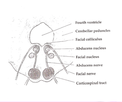 Axial section of the Brainstem (Pons) at the level of the Facial Colliculus (Central tegmental tract not labeled, but region is visible.) | |
| Details | |
| Identifiers | |
| Latin | Tractus tegmentalis centralis |
| NeuroNames | 2204 |
| TA98 | A14.1.05.325 |
| TA2 | 5869 |
| FMA | 83850 |
| Anatomical terms of neuroanatomy [edit on Wikidata] | |
The central tegmental tract[1] is a structure in the midbrain and pons.
It contains:
- ascending second-order axons projecting from the gustatory nucleus[citation needed] of nucleus solitarius[2]: 112 to the ventral posteromedial nucleus[2]: 473 of thalamus[2]: 112 (third-order neurons in turn project to the gustatory cortex)
- descending rubroolivary fibers projecting from the parvocellular red nucleus to the ipsilateral inferior olivary nucleus[2]: 292, 298 (which in turn projects to the contralateral cerebellum via olivocerebellar fibers[2]: 298 )
- ascending[2]: 306 reticulothalamic fibres[2]: 112 projecting from the medial zone nuclei of the reticular formation to the hypothalamus (to mediate autonomic nervous system response), and the intralaminar thalamic nuclei[2]: 306 (to mediate a startle response to pain[2]: 112 ).
Clinical significance
Lesion of the tract can cause palatal myoclonus, e.g. in myoclonic syndrome, in strokes of the posterior inferior cerebellar artery.
Additional Images
-
 Horizontal section through the lower part of the pons. The central tegmental tract is labeled #16.
 -
 Tractography showing central tegmental tract


References
- ^ Kamali A, Kramer LA, Butler IJ, Hasan KM. Diffusion tensor tractography of the somatosensory system in the human brainstem: initial findings using high isotropic spatial resolution at 3.0 T. Eur Radiol. 2009 19:1480-8. doi: 10.1007/s00330-009-1305-x. PMID 19189108
- ^ a b c d e f g h i Patestas, Maria A.; Gartner, Leslie P. (2016). A Textbook of Neuroanatomy (2nd ed.). Hoboken, New Jersey: Wiley-Blackwell. ISBN 978-1-118-67746-9.
External links
- Midbrain at Inferior Colliculus - IV Nucleus, Sectional Atlas
- Mid Pons at the Trigeminal Motor Nucleus, Sectional Atlas
- Neuroanatomy / plate12, Frank Willard
- v
- t
- e
Anatomy of the medulla
| Cranial nuclei |
| ||||
|---|---|---|---|---|---|
| Dorsal | |||||
| Ventral |
|
| Dorsal | |
|---|---|
| Ventral |
|
| Front | |
|---|---|
| Back |
| Authority control databases |
|
|---|


