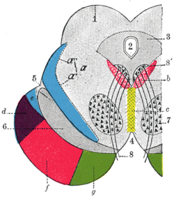
Temporopontine fibers
| Temporopontine fibers | |
|---|---|
 Coronal section through mid-brain.
| |
| Details | |
| Identifiers | |
| Latin | fibrae temporopontinae |
| NeuroNames | 1329 |
| Anatomical terms of neuroanatomy [edit on Wikidata] | |
The temporopontine fibers are corticopontine fibers projecting from the temporal lobe to the pontine nuclei. The temporopontine fibers are lateral to the cerebrospinal fibers; they originate in the temporal lobe and end in the pontine nuclei.
The fibers descend through the sublentiform part of the internal capsule.[1]
References
![]() This article incorporates text in the public domain from page 802 of the 20th edition of Gray's Anatomy (1918)
This article incorporates text in the public domain from page 802 of the 20th edition of Gray's Anatomy (1918)
- ^ Sinnatamby, Chummy S. (2011). Last's Anatomy (12th ed.). p. 461. ISBN 978-0-7295-3752-0.
- v
- t
- e
Anatomy of the midbrain
(Dorsal)
| Corpora quadrigemina | |||||
|---|---|---|---|---|---|
| Grey matter | |||||
| White matter |
|
(Ventral)
| Tegmentum |
| ||||||||||||||||
|---|---|---|---|---|---|---|---|---|---|---|---|---|---|---|---|---|---|
| Base |
| ||||||||||||||||
 | This neuroanatomy article is a stub. You can help Wikipedia by expanding it. |
- v
- t
- e


