DNAJA3
Protein-coding gene in the species Homo sapiens
| DNAJA3 | |||||||||||||||||||||||||||||||||||||||||||||||||||
|---|---|---|---|---|---|---|---|---|---|---|---|---|---|---|---|---|---|---|---|---|---|---|---|---|---|---|---|---|---|---|---|---|---|---|---|---|---|---|---|---|---|---|---|---|---|---|---|---|---|---|---|
 | |||||||||||||||||||||||||||||||||||||||||||||||||||
| Identifiers | |||||||||||||||||||||||||||||||||||||||||||||||||||
| Aliases | DNAJA3, HCA57, TID1, hTID-1, DnaJ heat shock protein family (Hsp40) member A3 | ||||||||||||||||||||||||||||||||||||||||||||||||||
| External IDs | OMIM: 608382; MGI: 1933786; HomoloGene: 36170; GeneCards: DNAJA3; OMA:DNAJA3 - orthologs | ||||||||||||||||||||||||||||||||||||||||||||||||||
| |||||||||||||||||||||||||||||||||||||||||||||||||||
| |||||||||||||||||||||||||||||||||||||||||||||||||||
| |||||||||||||||||||||||||||||||||||||||||||||||||||
| |||||||||||||||||||||||||||||||||||||||||||||||||||
| |||||||||||||||||||||||||||||||||||||||||||||||||||
| Wikidata | |||||||||||||||||||||||||||||||||||||||||||||||||||
| |||||||||||||||||||||||||||||||||||||||||||||||||||


DnaJ homolog subfamily A member 3, mitochondrial, also known as Tumorous imaginal disc 1 (TID1), is a protein that in humans is encoded by the DNAJA3 gene on chromosome 16.[5][6][7] This protein belongs to the DNAJ/Hsp40 protein family, which is known for binding and activating Hsp70 chaperone proteins to perform protein folding, degradation, and complex assembly.[6][7][8] As a mitochondrial protein, it is involved in maintaining membrane potential and mitochondrial DNA (mtDNA) integrity, as well as cellular processes such as cell movement, growth, and death.[6][7][9][10][11] Furthermore, it is associated with a broad range of diseases, including neurodegenerative diseases, inflammatory diseases, and cancers.[7][9][11][12]
Structure
As a member of the DNAJ/Hsp40 protein family, DNAJA3 contains a conserved DnaJ domain, which includes an HPD motif that interacts with Hsp70 to perform its cochaperone function.[6][7][8][9][10] The DnaJ domain is composed of tetrahelical regions containing a tripeptide of histidine, proline and aspartic acid situated between two helices. In addition, this protein contains a glycine/phenylalanine (G/F) rich linker region and a central cysteine-rich region similar to a zinc finger repeat, both characteristic of type I DnaJ molecular chaperones.[8][9][10] The mitochondrial targeting sequence at its N-terminal directs the localization of the protein to the mitochondrial matrix.[8][9][10]
DNAJA3 possesses two alternatively spliced forms: a long isoform of 43 kDa and a short isoform of 40 kDa.[6][7][9][12] The long isoform contains an additional 33 residues at its C-terminal compared to the short isoform, and this region is predicted to hinder the long isoform from regulating membrane potential.[7]
Function
DNAJA3 is a member of the DNAJ/Hsp40 protein family, which stimulates the ATPase activity of Hsp70 chaperones and plays critical roles in protein folding, degradation, and multiprotein complex assembly.[6][7][8] DNAJA3 localizes to the mitochondria, where it interacts with the mitochondrial Hsp70 chaperone (mtHsp70) to carry out the chaperone system.[6][7] This protein is crucial for maintaining a homogeneous distribution of mitochondrial membrane potential and the integrity of mtDNA. DNAJA3 homogenizes membrane potential through regulation of complex I aggregation, though the mechanism for maintaining mtDNA remains unknown.[7] These functions then allow DNAJA3 to mediate mitochondrial fission through DRP1 and, by extension, cellular processes such as cell movement, growth, proliferation, differentiation, senescence, and apoptosis.[6][7][9][10][11] However, though both isoforms of DNAJA3 are involved with cell survival, they are also observed to influence two opposing outcomes. The proapoptotic long isoform induces apoptosis by stimulating cytochrome C release and caspase activation in the mitochondria, whereas the antiapoptotic short isoform prevents cytochrome C release and, thus, apoptosis.[7][11] In neuromuscular junctions, only the short isoform clusters acetylcholine receptors for efficient synaptic transmission.[7] The two isoforms also differ in their specific mitochondrial localization, which may partially account for their different functions.[7][11]
Before localization to the mitochondria, DNAJA3 is transiently retained in the cytosol, where it can also interact with cytosolic proteins and possibly function to transport these proteins.[8][11]
Clinical significance
This protein is implicated in several cancers, including skin cancer, breast cancer, and colorectal cancer.[12] It is a key player in tumor suppression through interactions with oncogenic proteins, including ErbB2 and the p53 tumor suppressor protein.[6][8] Under hypoxic conditions, DNAJA3 may directly influence p53 complex assembly or modification, or indirectly ubiquitinylate p53 through ubiquitin ligases like MDM2. Moreover, both p53 and DNAJA3 must be present in the mitochondria in order to induce apoptosis in the cell.[8] In head and neck squamous cell carcinoma (HNSCC) cancer, DNAJA3 suppresses cell proliferation, anchorage-independent growth, cell motility, and cell invasion by attenuating EGFR and, downstream the signaling pathway, AKT.[12] Thus, treatments promoting DNAJA3 expression and function may greatly aid the elimination of tumors.[8]
Additionally, DNAJA3 is implicated in neurodegenerative diseases like Parkinson's disease by virtue of its key roles in chaperoning mitochondrial proteins and mediating mitochondrial morphology in conjunction with mtHsp70.[7][9] Another disease, psoriasis, is a chronic inflammatory skin disease that results from the absence of DNAJA3 activity, which then results in the activation of MK5, increased phosphorylation of HSP27, increased actin cytoskeleton organization, and hyperthickened skin.[11]
Interactions
DNAJA3 has been shown to interact with:
References
- ^ a b c ENSG00000103423 GRCh38: Ensembl release 89: ENSG00000276726, ENSG00000103423 – Ensembl, May 2017
- ^ a b c GRCm38: Ensembl release 89: ENSMUSG00000004069 – Ensembl, May 2017
- ^ "Human PubMed Reference:". National Center for Biotechnology Information, U.S. National Library of Medicine.
- ^ "Mouse PubMed Reference:". National Center for Biotechnology Information, U.S. National Library of Medicine.
- ^ Schilling B, De-Medina T, Syken J, Vidal M, Munger K (August 1998). "A novel human DnaJ protein, hTid-1, a homolog of the Drosophila tumor suppressor protein Tid56, can interact with the human papillomavirus type 16 E7 oncoprotein". Virology. 247 (1): 74–85. doi:10.1006/viro.1998.9220. PMID 9683573.
- ^ a b c d e f g h i "Entrez Gene: DNAJA3 DnaJ (Hsp40) homolog, subfamily A, member 3".
- ^ a b c d e f g h i j k l m n o p Ng, AC; Baird, SD; Screaton, RA (April 2014). "Essential role of TID1 in maintaining mitochondrial membrane potential homogeneity and mitochondrial DNA integrity". Molecular and Cellular Biology. 34 (8): 1427–37. doi:10.1128/mcb.01021-13. PMC 3993590. PMID 24492964.
- ^ a b c d e f g h i Ahn, BY; Trinh, DL; Zajchowski, LD; Lee, B; Elwi, AN; Kim, SW (25 February 2010). "Tid1 is a new regulator of p53 mitochondrial translocation and apoptosis in cancer". Oncogene. 29 (8): 1155–66. doi:10.1038/onc.2009.413. PMID 19935715.
- ^ a b c d e f g h Elwi, AN; Lee, B; Meijndert, HC; Braun, JE; Kim, SW (August 2012). "Mitochondrial chaperone DnaJA3 induces Drp1-dependent mitochondrial fragmentation". The International Journal of Biochemistry & Cell Biology. 44 (8): 1366–76. doi:10.1016/j.biocel.2012.05.004. PMID 22595283.
- ^ a b c d e f Trinh, DL; Elwi, AN; Kim, SW (October 2010). "Direct interaction between p53 and Tid1 proteins affects p53 mitochondrial localization and apoptosis". Oncotarget. 1 (6): 396–404. doi:10.18632/oncotarget.100902 (inactive 31 January 2024). PMC 3248115. PMID 21311096.
{{cite journal}}: CS1 maint: DOI inactive as of January 2024 (link) - ^ a b c d e f g h Choi, JH; Choi, DK; Sohn, KC; Kwak, SS; Suk, J; Lim, JS; Shin, I; Kim, SW; Lee, JH; Joe, CO (27 July 2012). "Absence of a human DnaJ protein hTid-1S correlates with aberrant actin cytoskeleton organization in lesional psoriatic skin". The Journal of Biological Chemistry. 287 (31): 25954–63. doi:10.1074/jbc.m111.313809. PMC 3406679. PMID 22692211.
- ^ a b c d Chen, CY; Chiou, SH; Huang, CY; Jan, CI; Lin, SC; Hu, WY; Chou, SH; Liu, CJ; Lo, JF (November 2009). "Tid1 functions as a tumour suppressor in head and neck squamous cell carcinoma". The Journal of Pathology. 219 (3): 347–55. doi:10.1002/path.2604. PMID 19681071. S2CID 23405415.
- ^ a b Sarkar S, Pollack BP, Lin KT, Kotenko SV, Cook JR, Lewis A, Pestka S (December 2001). "hTid-1, a human DnaJ protein, modulates the interferon signaling pathway". J. Biol. Chem. 276 (52): 49034–42. doi:10.1074/jbc.M103683200. PMID 11679576.
- ^ Trentin GA, Yin X, Tahir S, Lhotak S, Farhang-Fallah J, Li Y, Rozakis-Adcock M (April 2001). "A mouse homologue of the Drosophila tumor suppressor l(2)tid gene defines a novel Ras GTPase-activating protein (RasGAP)-binding protein". J. Biol. Chem. 276 (16): 13087–95. doi:10.1074/jbc.M009267200. PMID 11116152.
Further reading
- Maruyama K, Sugano S (1994). "Oligo-capping: a simple method to replace the cap structure of eukaryotic mRNAs with oligoribonucleotides". Gene. 138 (1–2): 171–4. doi:10.1016/0378-1119(94)90802-8. PMID 8125298.
- Suzuki Y, Yoshitomo-Nakagawa K, Maruyama K, et al. (1997). "Construction and characterization of a full length-enriched and a 5'-end-enriched cDNA library". Gene. 200 (1–2): 149–56. doi:10.1016/S0378-1119(97)00411-3. PMID 9373149.
- Syken J, De-Medina T, Münger K (1999). "TID1, a human homolog of the Drosophila tumor suppressor l(2)tid, encodes two mitochondrial modulators of apoptosis with opposing functions". Proc. Natl. Acad. Sci. U.S.A. 96 (15): 8499–504. Bibcode:1999PNAS...96.8499S. doi:10.1073/pnas.96.15.8499. PMC 17545. PMID 10411904.
- Shinohara M, Gasior SL, Bishop DK, Shinohara A (2000). "Tid1/Rdh54 promotes colocalization of rad51 and dmc1 during meiotic recombination". Proc. Natl. Acad. Sci. U.S.A. 97 (20): 10814–9. Bibcode:2000PNAS...9710814S. doi:10.1073/pnas.97.20.10814. PMC 27106. PMID 11005857.
- Trentin GA, Yin X, Tahir S, et al. (2001). "A mouse homologue of the Drosophila tumor suppressor l(2)tid gene defines a novel Ras GTPase-activating protein (RasGAP)-binding protein". J. Biol. Chem. 276 (16): 13087–95. doi:10.1074/jbc.M009267200. PMID 11116152.
- Ohtsuka K, Hata M (2001). "Mammalian HSP40/DNAJ homologs: cloning of novel cDNAs and a proposal for their classification and nomenclature". Cell Stress Chaperones. 5 (2): 98–112. doi:10.1379/1466-1268(2000)005<0098:mhdhco>2.0.co;2 (inactive 2024-02-01). ISSN 1466-1268. PMC 312896. PMID 11147971.
{{cite journal}}: CS1 maint: DOI inactive as of February 2024 (link) - Sarkar S, Pollack BP, Lin KT, et al. (2002). "hTid-1, a human DnaJ protein, modulates the interferon signaling pathway". J. Biol. Chem. 276 (52): 49034–42. doi:10.1074/jbc.M103683200. PMID 11679576.
- Yin X, Rozakis-Adcock M (2002). "Genomic organization and expression of the human tumorous imaginal disc (TID1) gene". Gene. 278 (1–2): 201–10. doi:10.1016/S0378-1119(01)00720-X. PMID 11707338.
- Cheng H, Cenciarelli C, Shao Z, et al. (2002). "Human T cell leukemia virus type 1 Tax associates with a molecular chaperone complex containing hTid-1 and Hsp70". Curr. Biol. 11 (22): 1771–5. doi:10.1016/S0960-9822(01)00540-1. PMID 11719219. S2CID 17461974.
- Cheng H, Cenciarelli C, Tao M, et al. (2002). "HTLV-1 Tax-associated hTid-1, a human DnaJ protein, is a repressor of Ikappa B kinase beta subunit". J. Biol. Chem. 277 (23): 20605–10. doi:10.1074/jbc.M201204200. PMID 11927590.
- Wang Y, Han KJ, Pang XW, et al. (2002). "Large scale identification of human hepatocellular carcinoma-associated antigens by autoantibodies". J. Immunol. 169 (2): 1102–9. doi:10.4049/jimmunol.169.2.1102. PMID 12097419.
- Sasaki S, Nakamura T, Arakawa H, et al. (2002). "Isolation and characterization of a novel gene, hRFI, preferentially expressed in esophageal cancer". Oncogene. 21 (32): 5024–30. doi:10.1038/sj.onc.1205627. PMID 12118383.
- Strausberg RL, Feingold EA, Grouse LH, et al. (2003). "Generation and initial analysis of more than 15,000 full-length human and mouse cDNA sequences". Proc. Natl. Acad. Sci. U.S.A. 99 (26): 16899–903. Bibcode:2002PNAS...9916899M. doi:10.1073/pnas.242603899. PMC 139241. PMID 12477932.
- Syken J, Macian F, Agarwal S, et al. (2003). "TID1, a mammalian homologue of the drosophila tumor suppressor lethal(2) tumorous imaginal discs, regulates activation-induced cell death in Th2 cells". Oncogene. 22 (30): 4636–41. doi:10.1038/sj.onc.1206569. PMID 12879007.
- Rodriguez M, Yu X, Chen J, Songyang Z (2004). "Phosphopeptide binding specificities of BRCA1 COOH-terminal (BRCT) domains". J. Biol. Chem. 278 (52): 52914–8. doi:10.1074/jbc.C300407200. PMID 14578343.
- Ota T, Suzuki Y, Nishikawa T, et al. (2004). "Complete sequencing and characterization of 21,243 full-length human cDNAs". Nat. Genet. 36 (1): 40–5. doi:10.1038/ng1285. PMID 14702039.
- Lo JF, Hayashi M, Woo-Kim S, et al. (2004). "Tid1, a cochaperone of the heat shock 70 protein and the mammalian counterpart of the Drosophila tumor suppressor l(2)tid, is critical for early embryonic development and cell survival". Mol. Cell. Biol. 24 (6): 2226–36. doi:10.1128/MCB.24.6.2226-2236.2004. PMC 355836. PMID 14993262.
- Edwards KM, Münger K (2004). "Depletion of physiological levels of the human TID1 protein renders cancer cell lines resistant to apoptosis mediated by multiple exogenous stimuli". Oncogene. 23 (52): 8419–31. doi:10.1038/sj.onc.1207732. PMID 15156195.
- Colland F, Jacq X, Trouplin V, et al. (2004). "Functional proteomics mapping of a human signaling pathway". Genome Res. 14 (7): 1324–32. doi:10.1101/gr.2334104. PMC 442148. PMID 15231748.
- v
- t
- e
PDB gallery
-
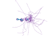 2ctt: Solution structure of zinc finger domain from human DnaJ subfamily A member 3
 -
 2dn9: Solution structure of J-domain from the DnaJ homolog, human Tid1 protein


